Your Plant stem cross section images are available. Plant stem cross section are a topic that is being searched for and liked by netizens today. You can Find and Download the Plant stem cross section files here. Get all royalty-free vectors.
If you’re searching for plant stem cross section images information related to the plant stem cross section topic, you have visit the ideal site. Our site always provides you with suggestions for seeing the maximum quality video and picture content, please kindly surf and locate more informative video content and images that fit your interests.
Plant Stem Cross Section. New parts of the stem. If you were to look carefully at the cross section of a stem, you would find several layers inside, each of which has a different job. Three terms, epidermis, cortex and pith are used to broadly describe the distribution of tissues across the stem. Inferior ovary, rose , rosaceae.
 stem Description, Facts, & Types Britannica From britannica.com
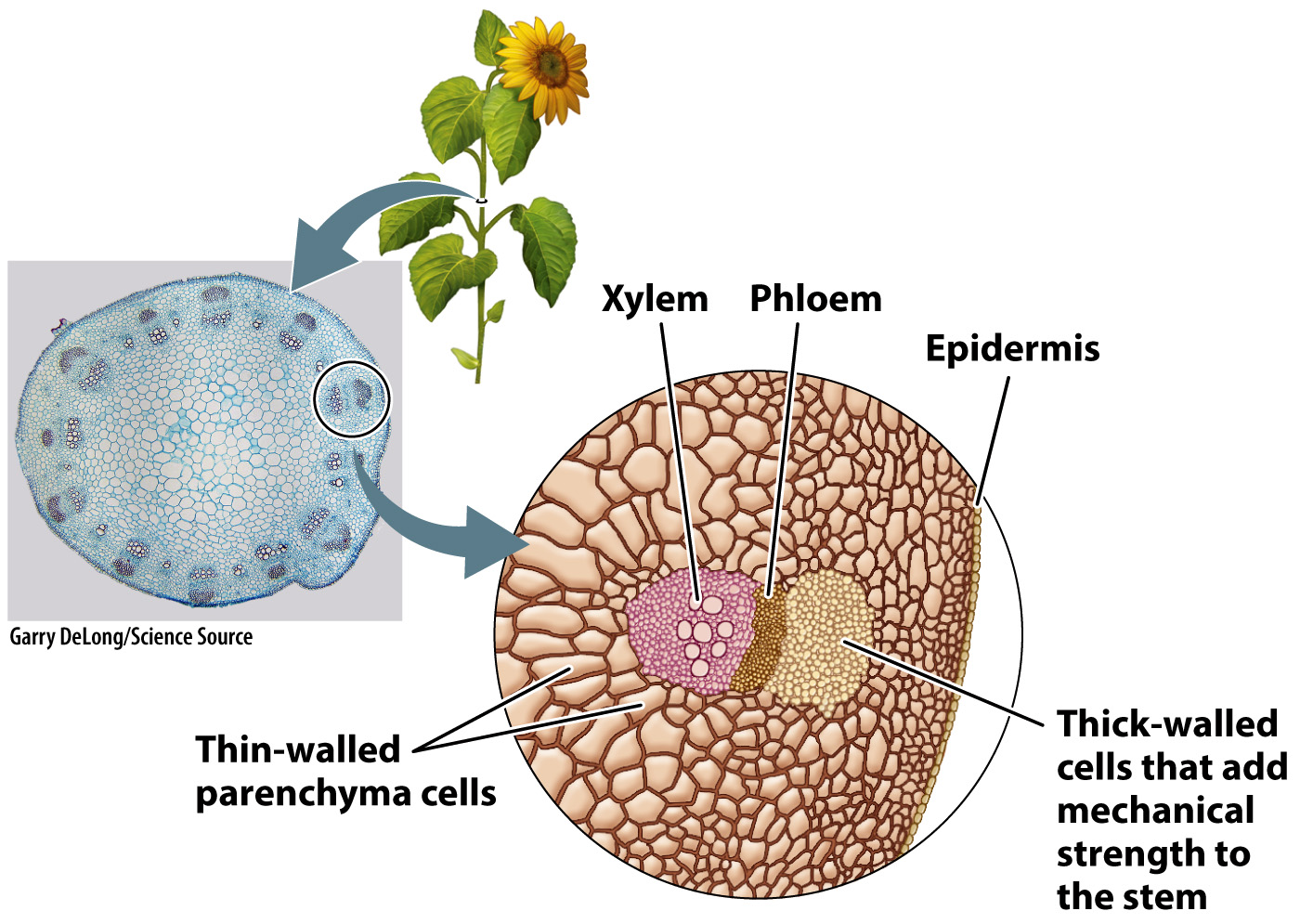
Dicot vascular bundles of xylem and phloem are arranged in a ring. In a cross section, the pith may be rounded, triangular or star shaped. Xylem cells are dead, elongated, and hollow. Protective covering of the stem. The large spring xylem cells. Scattered throughout the stem in bundles.
Dicot stems with primary growth have pith in the center, with vascular bundles forming a distinct ring visible when the stem is viewed in cross section.
Protective covering of the stem. In a cross section, the pith may be rounded, triangular or star shaped. Xylem cells are dead, elongated, and hollow. Three terms, epidermis, cortex and pith are used to broadly describe the distribution of tissues across the stem. The large spring xylem cells. A very young stem of leonurus sibiricus of family labiatae should be selected, because secondary growth commences unusually early in this plant.
 Source: cronodon.com
Monocot stem cross section 4. Download this stem of lycopodium microscopic cross section cut of a plant stem photo now. Study of flowers, leaves and inflorescences of dicotyledon plants, drawing. Then, you can stain the cross section of plant stems with methylene blue or eosin y (or both) and start looking them under the microscope. Xylem cells are dead, elongated, and hollow.
 Source: oercommons.org
For studying the internal structure of a typical dicot stem, the stem cross section of a young sunflower or cucurbita is taken. Monocot leaf cross section 3. Plant organ cross sections 2. Outer layer of the stem. Cross section of dicot stem seen under a microscope.
 Source: courses.lumenlearning.com
Epidermis of a leaf of redgal (morinda royoc, an angiosperm). Plant stems perform two basic functions: The outside of the stem is covered with an. If you were to look carefully at the cross section of a stem, you would find several layers inside, each of which has a different job. Epidermis of a leaf of redgal (morinda royoc, an angiosperm).
 Source: pinterest.co.uk
Browse 2,069 stem cross section stock photos and images available, or search for plant cross section or ranunculus cross section to find more great stock photos and pictures. Monocot leaf cross section 3. A thin transverse section of the young dicot root of gram, sunflower or pea reveals the following structures under the microscope: New parts of the stem. The large spring xylem cells.
 Source: macmillanhighered.com
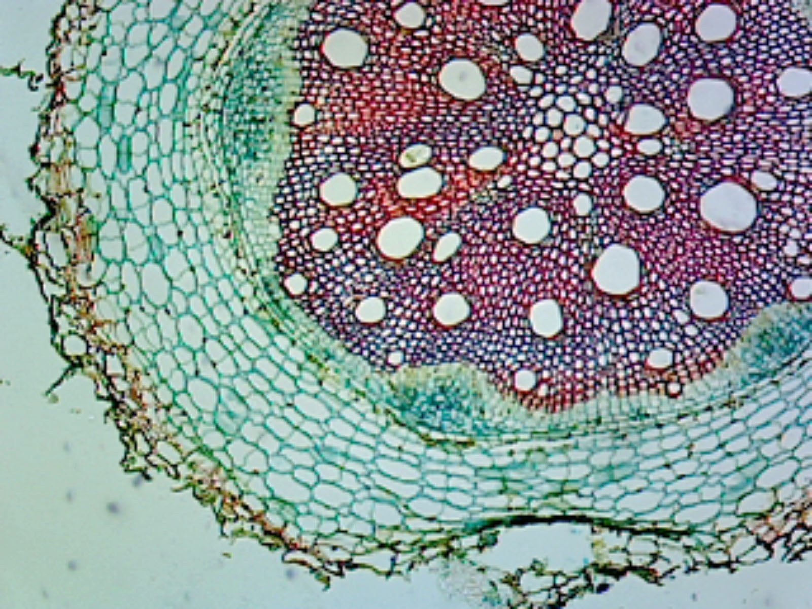
Dicot root cross section 8. Cotton (gossypium spec.), cross section of a stem of a cotton plant, 100 x, detail of xylem, phloem and cambium cross section of vascular tissue system of a plant. Cally on xylem and phloem of plant stems, branches and roots. The stem is square in. New parts of the stem.
 Source: pinterest.com.au
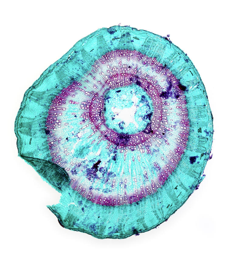
They support the leaves and flowers and they carry water and food from place to place within the plant. A typical stem is cylindrical and may be soft ( herbaceous) or woody. The arrangement of the vascular tissues varies widely among plant species. Cut a piece about 1cm long (make sure its longer than the depth of the nut of your hand microtome. Xylem growth makes the “annual rings” used to tell a tree’s age.
 Source: fineartamerica.com
The dermal tissue of aquatic plants stems may lack the waterproofing found in aerial stems. Monocot root cross section 5. In a cross section, the pith may be rounded, triangular or star shaped. A thin transverse section of the young dicot root of gram, sunflower or pea reveals the following structures under the microscope: Although young children may not be quite ready to.

It is usually branched and leafy. Photo about microscopic view of a plant stem cross cut section under the scientific microscope. Browse 237 dicot stem cross section stock photos and images available, or start a new search to explore more stock photos and images. Dicot stem cross section 7. The xylem is responsible for keeping a plant hydrated by transporting water upward from the roots.

Xylem cells are dead, elongated, and hollow. Dicot root cross section dicot root diagram reveals internal structure of dicot root. The large spring xylem cells. Central part of the stem. The outside of the stem is covered with an.
 Source: britannica.com
Download this stem of lycopodium microscopic cross section cut of a plant stem photo now. Central part of the stem. Dicot leaf cross section 6. The point at which a leaf joins the stem is called the node. Cross section of a polyarch root of aeglopsis chevalieri, a plant in the citrus family (rutaceae) of angiosperms.
 Source: pinterest.com
As we just went over, there are many different components of the shoot system. Protective covering of the stem. Although young children may not be quite ready to. Central part of the stem. Plant organ cross sections 2.

The arrangement of the vascular tissues varies widely among plant species. In a cross section, the pith may be rounded, triangular or star shaped. Dicot stems with primary growth have pith in the center, with vascular bundles forming a distinct ring visible when the stem is viewed in cross section. The large spring xylem cells. Plant stems perform two basic functions:
 Source: researchgate.net
Xylem growth makes the “annual rings” used to tell a tree’s age. Three terms, epidermis, cortex and pith are used to broadly describe the distribution of tissues across the stem. It forms a single and the outermost layer of the stem. Stem pith is used in plant identification. Cut a piece about 1cm long (make sure its longer than the depth of the nut of your hand microtome.
 Source: pinterest.com
Protective covering of the stem. Three terms, epidermis, cortex and pith are used to broadly describe the distribution of tissues across the stem. Dicot root cross section dicot root diagram reveals internal structure of dicot root. Dicot stems with primary growth have pith in the center, with vascular bundles forming a distinct ring visible when the stem is viewed in cross section. The xylem is responsible for keeping a plant hydrated by transporting water upward from the roots.
 Source: fineartamerica.com
The arrangement of the vascular tissues varies widely among plant species. Dicot root cross section 8. In this lesson, we will focus on the stem. Dicot stem cross section 7. Epidermis of a leaf of redgal (morinda royoc, an angiosperm).
 Source: pinterest.fr
Plant organ cross sections 1. Each vascular bundle consists of three parts. Transverse sections are taken and stained suitably for the internal structure. Let them float on the surface of the water. The dermal tissue of aquatic plants stems may lack the waterproofing found in aerial stems.

A thin transverse section of the young dicot root of gram, sunflower or pea reveals the following structures under the microscope: Xylem growth makes the “annual rings” used to tell a tree’s age. Browse 237 dicot stem cross section stock photos and images available, or start a new search to explore more stock photos and images. Browse 2,069 stem cross section stock photos and images available, or search for plant cross section or ranunculus cross section to find more great stock photos and pictures. It forms a single and the outermost layer of the stem.
 Source: walmart.com
Protective covering of the stem. A thin transverse section of the young dicot root of gram, sunflower or pea reveals the following structures under the microscope: Browse 237 dicot stem cross section stock photos and images available, or start a new search to explore more stock photos and images. In woody dicot plants, the rings grow to make a complete ring around the stem. Three terms, epidermis, cortex and pith are used to broadly describe the distribution of tissues across the stem.
This site is an open community for users to do sharing their favorite wallpapers on the internet, all images or pictures in this website are for personal wallpaper use only, it is stricly prohibited to use this wallpaper for commercial purposes, if you are the author and find this image is shared without your permission, please kindly raise a DMCA report to Us.
If you find this site value, please support us by sharing this posts to your preference social media accounts like Facebook, Instagram and so on or you can also bookmark this blog page with the title plant stem cross section by using Ctrl + D for devices a laptop with a Windows operating system or Command + D for laptops with an Apple operating system. If you use a smartphone, you can also use the drawer menu of the browser you are using. Whether it’s a Windows, Mac, iOS or Android operating system, you will still be able to bookmark this website.


