Your Plant under microscope images are available. Plant under microscope are a topic that is being searched for and liked by netizens now. You can Get the Plant under microscope files here. Download all free images.
If you’re searching for plant under microscope pictures information linked to the plant under microscope topic, you have come to the ideal blog. Our site always gives you suggestions for viewing the maximum quality video and image content, please kindly surf and locate more informative video articles and images that match your interests.
Plant Under Microscope. Plant cell diagram under electron microscope. The cell also appears green in color due to the chlorophyll pigment within the chloroplasts. The cell wall is distinctly visible around each cell. To prepare plant cells for viewing under a microscope place a drop of iodine onto a clean glass slide iodine is used to stain dye a cell and make it easier to see.
 Pin on NanoTechnia From pinterest.ie
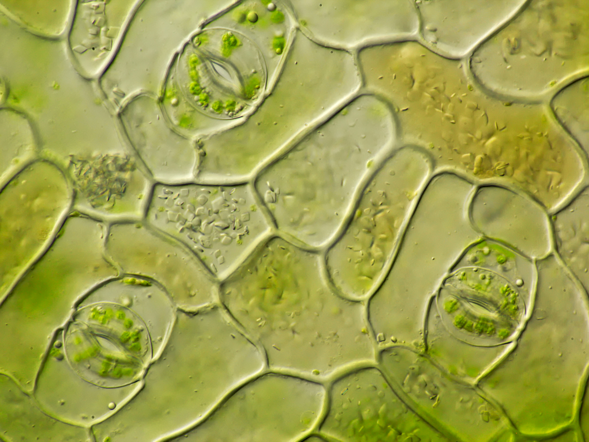
Other organelles may also be seen, depending on. Ad mvx10 macroview microscope for efficient, bright fluorescence imaging. Microscopic photo of a plant cell with red and green colors and cell textures. Similarly, what does a plant cell look like under a microscope? Under the microscope, plant cells are seen as large rectangular interlocking blocks. Plant stomata under the microscope on the outer layer of the leaf of a plant are microscopic holes called stomata.
What does a plant cell look like under a microscope?
Studying cell tissues from an onion peel is a great exercise in using light microscopes and learning about plant cells, since onion cells are highly visible under a microscope, especially when stained correctly. To prepare plant cells for viewing under a microscope place a drop of iodine onto a clean glass slide iodine is used to stain dye a cell and make it easier to see. The five main parts are the roots, the leaves, the stem, the flower, and the seed. Using a camera or cell phone, images of microscope slide contents allow students to label plant parts and engage in discussions with peers. Under a microscope, plant cells from the same source will have a uniform size and shape.beneath a plant cell's cell wall is a cell membrane. Plant cell diagram the plant cell is rectangular and comparatively larger than the animal cell.
 Source: pinterest.se
Plant cells under the microscope. Under a microscope, plant cells from the same source will have a uniform size and shape.beneath a plant cell's cell wall is a cell membrane. Cross section of the stem and thallus of a fungus under a microscope, drawing. Under the microscope, plant cells are seen as large rectangular interlocking blocks. Ad mvx10 macroview microscope for efficient, bright fluorescence imaging.
 Source: ar.pinterest.com
Leaf with chloroplast from pondweed , hydrocharitaceae, seen under a microscope. Similarly, what does a plant cell look like under a microscope? When seen under a microscope, a plant cell is somewhat rectangular in shape and displays a double membrane which is more rigid than that of an animal cell. Leaf cross section under the microscope Look at the leaf down a microscope and see if you can identify the small green chloroplasts.
 Source: pinterest.com
Looking at chloroplasts under the microscope. Other organelles may also be seen, depending on. The cell wall is somewhat thick and is seen rightly when stained. In this activity, students section plant material and prepare specimens to view under a brightfield microscope. To prepare plant cells for viewing under a microscope place a drop of iodine onto a clean glass slide iodine is used to stain dye a cell and make it easier to see.
 Source: pinterest.com
Similarly, what does a plant cell look like under a microscope? Tracking individual cells in the growing tips of roots and shoots helps scientists understand how these precursor stem cells differentiate into specialised plant organs like. Similarly, what does a plant cell look like under a microscope? While the slcu research team of alexander jones is developing biosensors for confocal microscopes that enable plant scientists to track in real time the molecules behind plant growth. Cross section of the stem and thallus of a fungus under a microscope, drawing.
 Source: pinterest.com
In this activity, students section plant material and prepare specimens to view under a brightfield microscope. In this activity, students section plant material and prepare specimens to view under a brightfield microscope. Similarly, what does a plant cell look like under a microscope? Leaf with chloroplast from pondweed , hydrocharitaceae, seen under a microscope. Ad mvx10 macroview microscope for efficient, bright fluorescence imaging.

Under the microscope, plant cells are seen as large rectangular interlocking blocks. By looking at the microscopic structures of different parts of the plant parts, we can learn how the plant function at the cellular level. Red blood cells (rbcs) as seen under the microscope in isotonic, hypotonic and hypertonic solutions. For our science microscope activity, we will be examining the stem of a plant under the microscope. Observe fern spore under a microscope the material you need.
 Source: hempfy.com
Looking at chloroplasts under the microscope. Each part has its unique job to keep the whole plant healthy. Plant cell diagram under electron microscope. See more ideas about microscope, simple green, microscopic. Under high magnification, students can differentiate between closed and open stomata.
 Source: pinterest.ie
Place a single leaf on a microscope slide, add a drop of water and a cover slip. The cell also appears green in color due to the chlorophyll pigment within the chloroplasts. Under the microscope, plant cells are seen as large rectangular interlocking blocks. For our science microscope activity, we will be examining the stem of a plant under the microscope. Editible eps vector file the animal cell diagram vector etsy in 2021 animal cell cell diagram plant cell diagram.
 Source: saxon.com.au
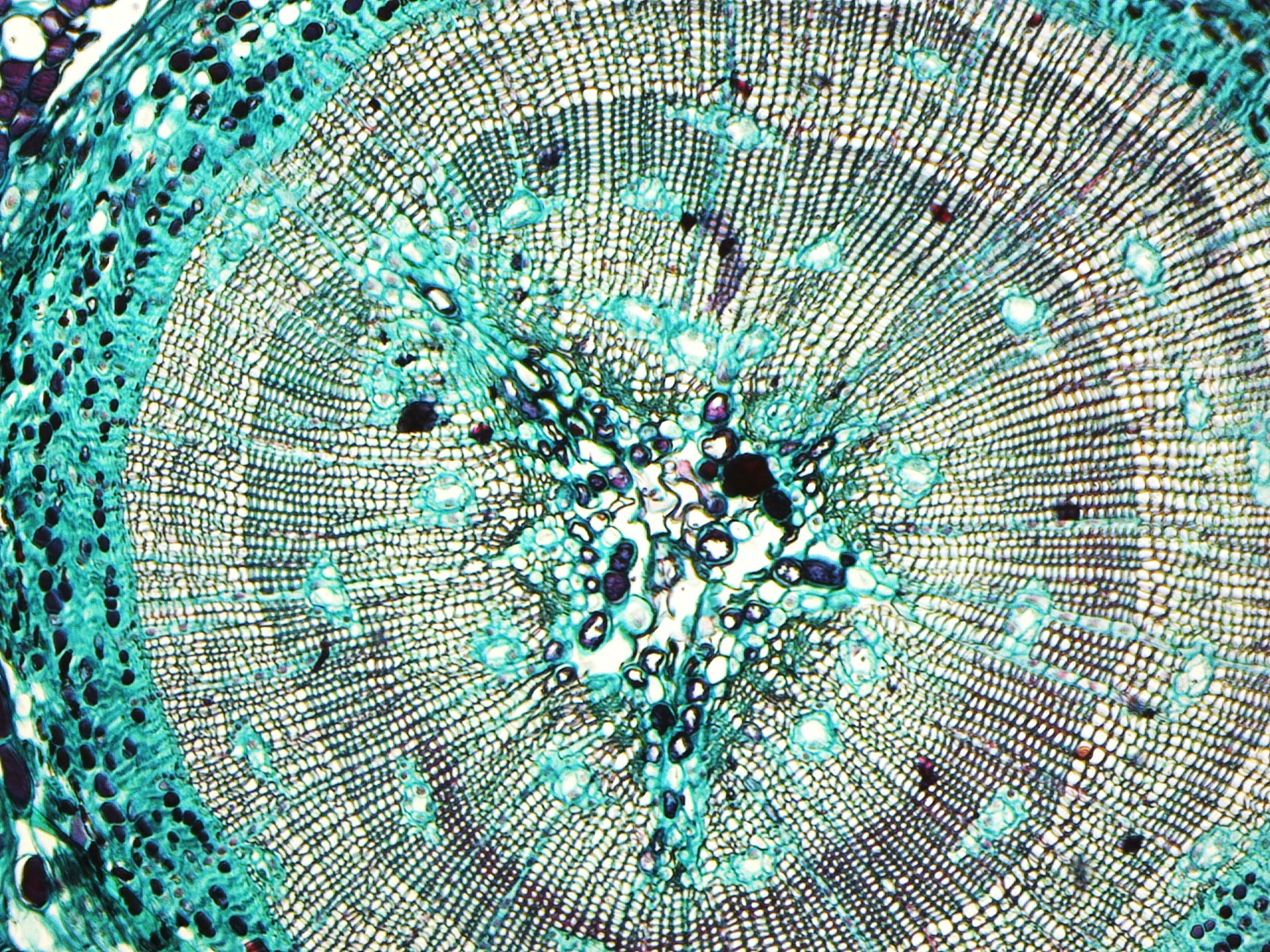
This can prove to be a very interesting as well as educational activity for students and children alike especially those from high school and elementary. Plant stem section under the microscope detail. Compare and contrast animal and plant cells and be able to distinguish each type under the microscope. In this activity, students section plant material and prepare specimens to view under a brightfield microscope. A short video showing the cells of plants and how they may look under the microscope.
 Source: cannsociety.com
What does a plant cell look like under a microscope? Similarly, what does a plant cell look like under a microscope? While the slcu research team of alexander jones is developing biosensors for confocal microscopes that enable plant scientists to track in real time the molecules behind plant growth. Editible eps vector file the animal cell diagram vector etsy in 2021 animal cell cell diagram plant cell diagram. Under a microscope, plant cells from the same source will have a uniform size and shape.beneath a plant cell's cell wall is a cell membrane.
 Source: microscopyofnature.com
Each part has its unique job to keep the whole plant healthy. 40x 400x compound monocularbiological microscope45 degree angled headelectric lightedbeginner slides plant cell things under a microscope plant cell picture. The diagram is very clear and labeled. Observe fern spore under a microscope the material you need. The cell wall is somewhat thick and is seen rightly when stained.
 Source: pinterest.co.uk
Plant cell from a leaf is large and occupies about 80 of the cell volume the photograph shown below details chloroplast structure as viewed with a transmission electron microscope. Angelo on january 3, 2022. Under high magnification, students can differentiate between closed and open stomata. Nisa on december 15, 2021. Under the microscope, plant cells are seen as large rectangular interlocking blocks.
 Source: pulpbits.net
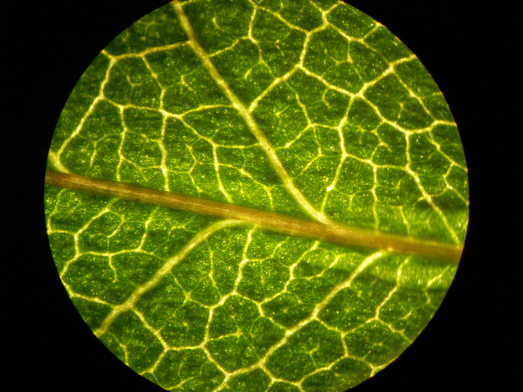
See more ideas about microscope, simple green, microscopic. Similarly, what does a plant cell look like under a microscope? Plant cell diagram under electron microscope. Leaf cross section under the microscope A plant is made up of several different parts.
 Source: pinterest.dk
Angelo on january 3, 2022. Ad mvx10 macroview microscope for efficient, bright fluorescence imaging. What does a plant cell look like under a microscope? Scanning electron microscope micrograph showing granules of corn starch, at a magnification of 400x, 2016. Plant cells under the microscope.

Plant cell diagram under electron microscope. Leaf cross section under the microscope The cell also appears green in color due to the chlorophyll pigment within the chloroplasts. The black sorus is riper than the white one. Other organelles may also be seen, depending on.
 Source: pinterest.com
The vegetative structure or plant body of spirogyra is known as thallus. Nisa on december 15, 2021. Plant stomata under the microscope on the outer layer of the leaf of a plant are microscopic holes called stomata. Typically, the stomata are bean shaped and will appear denser (darker) under the microscope. Pay attention to the color of the sori.
 Source: pinterest.com
Plant cell diagram under electron microscope. Red blood cells (rbcs) as seen under the microscope in isotonic, hypotonic and hypertonic solutions. The vegetative structure or plant body of spirogyra is known as thallus. The diagram is very clear and labeled. Plant cell diagram the plant cell is rectangular and comparatively larger than the animal cell.
 Source: piqueen.com
Compare and contrast animal and plant cells and be able to distinguish each type under the microscope. Ad mvx10 macroview microscope for efficient, bright fluorescence imaging. Red blood cells (rbcs) as seen under the microscope in isotonic, hypotonic and hypertonic solutions. Ad mvx10 macroview microscope for efficient, bright fluorescence imaging. Similarly, what does a plant cell look like under a microscope?
This site is an open community for users to submit their favorite wallpapers on the internet, all images or pictures in this website are for personal wallpaper use only, it is stricly prohibited to use this wallpaper for commercial purposes, if you are the author and find this image is shared without your permission, please kindly raise a DMCA report to Us.
If you find this site good, please support us by sharing this posts to your preference social media accounts like Facebook, Instagram and so on or you can also save this blog page with the title plant under microscope by using Ctrl + D for devices a laptop with a Windows operating system or Command + D for laptops with an Apple operating system. If you use a smartphone, you can also use the drawer menu of the browser you are using. Whether it’s a Windows, Mac, iOS or Android operating system, you will still be able to bookmark this website.


