Your Plantar and dorsal foot images are available in this site. Plantar and dorsal foot are a topic that is being searched for and liked by netizens now. You can Get the Plantar and dorsal foot files here. Find and Download all royalty-free photos and vectors.
If you’re searching for plantar and dorsal foot images information connected with to the plantar and dorsal foot topic, you have pay a visit to the ideal blog. Our website frequently gives you suggestions for seeking the maximum quality video and image content, please kindly surf and find more enlightening video articles and graphics that match your interests.
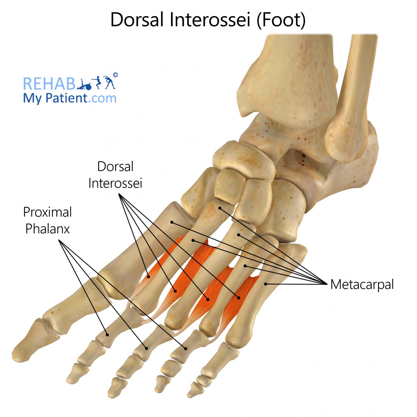
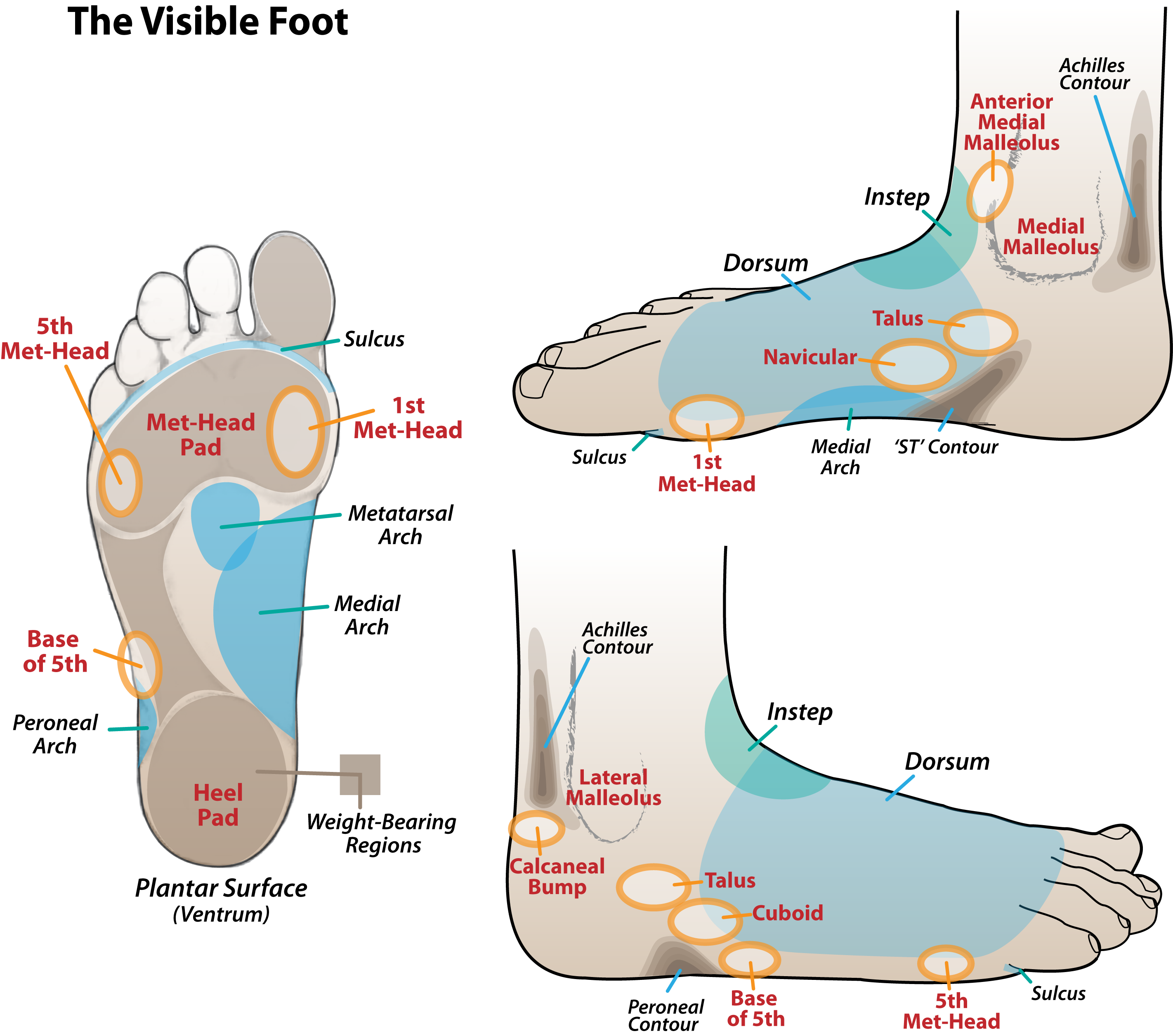
Plantar And Dorsal Foot. The top of the foot is called the dorsum of the foot. Borrowed from latin plantāre, present active infinitive of plantō. Each arises from a single metatarsal. Plantar interossei are invested by the plantar interosseous fascia and accompanied by the dorsal interossei muscles.
 Dorsal muscles of the foot Anatomy and function Kenhub From kenhub.com
The force was higher on the plantar. As adjectives the difference between dorsal and plantar is that dorsal is (anatomy) with respect to, or concerning the side in which the backbone is located, or the analogous side of an invertebrate while plantar is pertaining to the bottom surface ( sole ) of the foot, as with plantar warts compare palmar. Dorsiflexion occurs in both ankle joint and wrist joint. There are three plantar interossei, which are located between the metatarsals. Anterior tibial artery, via dorsalis pedis and dorsal metatarsal arteries Plantar and dorsal foot anatomy.
The plantar ligaments are stronger than those on the dorsal side (figure 12 & 13).
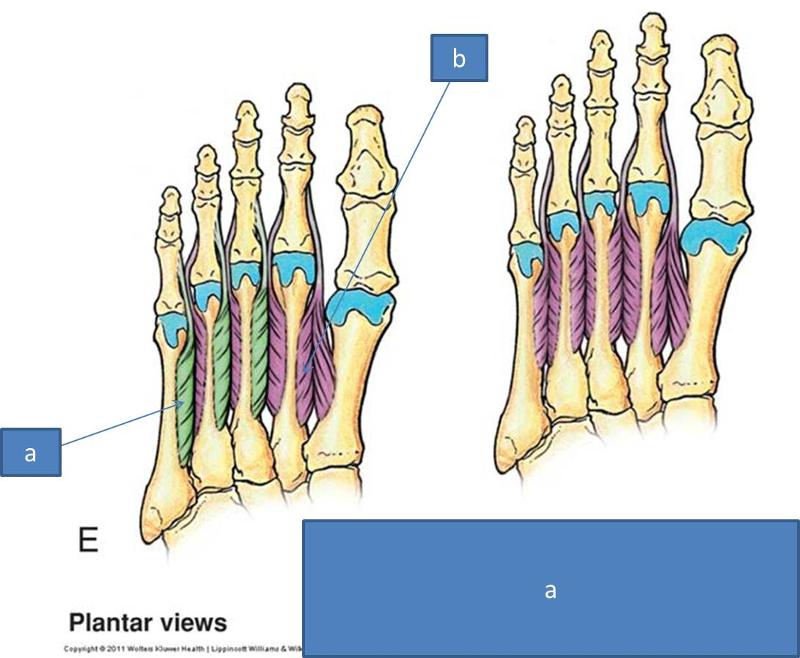
Plantar flexion is the lifting of the heel of the foot from the ground or pointing the toes downward. Anterior tibial artery, via dorsalis pedis and dorsal metatarsal arteries Plantar interossei ( 3) dorsal interossei ( 4) plantar interossei origin. Plantar interossei are smaller than their dorsal counterparts and lie below the metatarsal bones. The plantar interossei have a unipennate morphology, while the dorsal interossei are bipennate. But, plantar flexion only occurs in the ankle joint.
 Source: researchgate.net
Plantar interossei are smaller than their dorsal counterparts and lie below the metatarsal bones. The lisfranc ligament is a strong band of tissue that connects the medial cuneiform to the base of the second metatarsal. Request pdf | plantar and dorsal foot loading measurements in patients after rotationplasty | the present study investigated the plantar and dorsal foot loading patterns inside the prosthesis of. The dorsal loading area was smaller than the plantar area (p=0.003). Note the dorsal surfaces of the body, muzzle, feet.
 Source: kenhub.com
These ligaments prevent the joints of the midfoot from moving much, and as such provide considerable stability to the arch of the foot. During dorsiflexion, the angle between leg and the dorsum of the foot is decreased. If you’ve ever had a plantar wart, then you’ve had a wart on the sole of your foot (ouch!). Plantar arterial supply posterior tibial artery gives off its calcaneal branch then divides into the medial and lateral plantar arteries medial plantar artery branch of. (anatomy) relating to the sole of the foot.
 Source: dedicated11.blogspot.com
Flexor digiti minimi brevis innervation. Blood supply the vascularization of dorsal interossei muscles comes from several small arteries in the foot ; As adjectives the difference between dorsal and plantar is that dorsal is (anatomy) with respect to, or concerning the side in which the backbone is located, or the analogous side of an invertebrate while plantar is pertaining to the bottom surface ( sole ) of the foot, as with plantar warts compare palmar. The base or bottom (or pad) of the foot. The lisfranc ligament is a strong band of tissue that connects the medial cuneiform to the base of the second metatarsal.
 Source: rehabmypatient.com
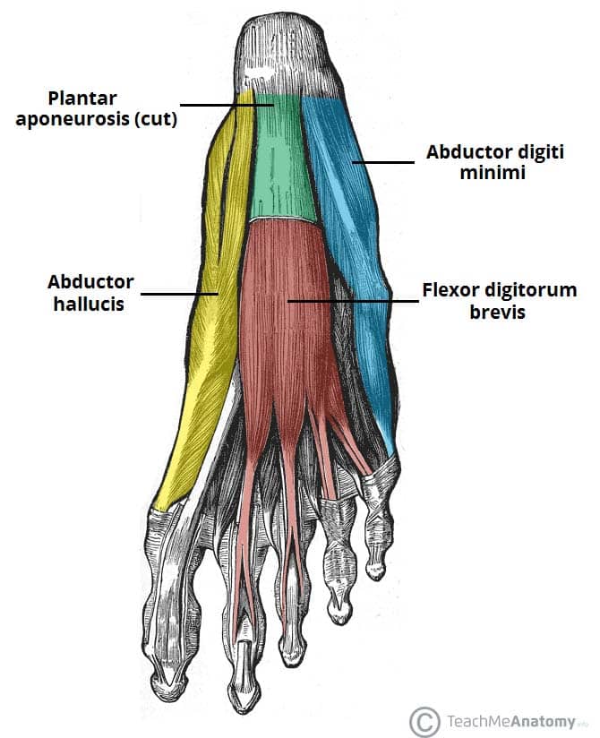
In humans the sole of the foot is anatomically referred to as the plantar aspect. Medial process of calcaneal tuberosity. The plantar and dorsal interossei comprise the fourth and final plantar muscle layer. (anatomy) relating to the sole of the foot. Each arises from a single metatarsal.
 Source: learnmuscles.com
Plantar arterial supply posterior tibial artery gives off its calcaneal branch then divides into the medial and lateral plantar arteries medial plantar artery branch of. In anatomy, the sole of the foot is called the plantar surface. The dorsal loading area was smaller than the plantar area (p=0.003). Dorsiflexion and plantar flexion are two motions of the body, decreasing the angle between two anatomical parts of the body. The sole is the bottom of the foot.
 Source: pinterest.com
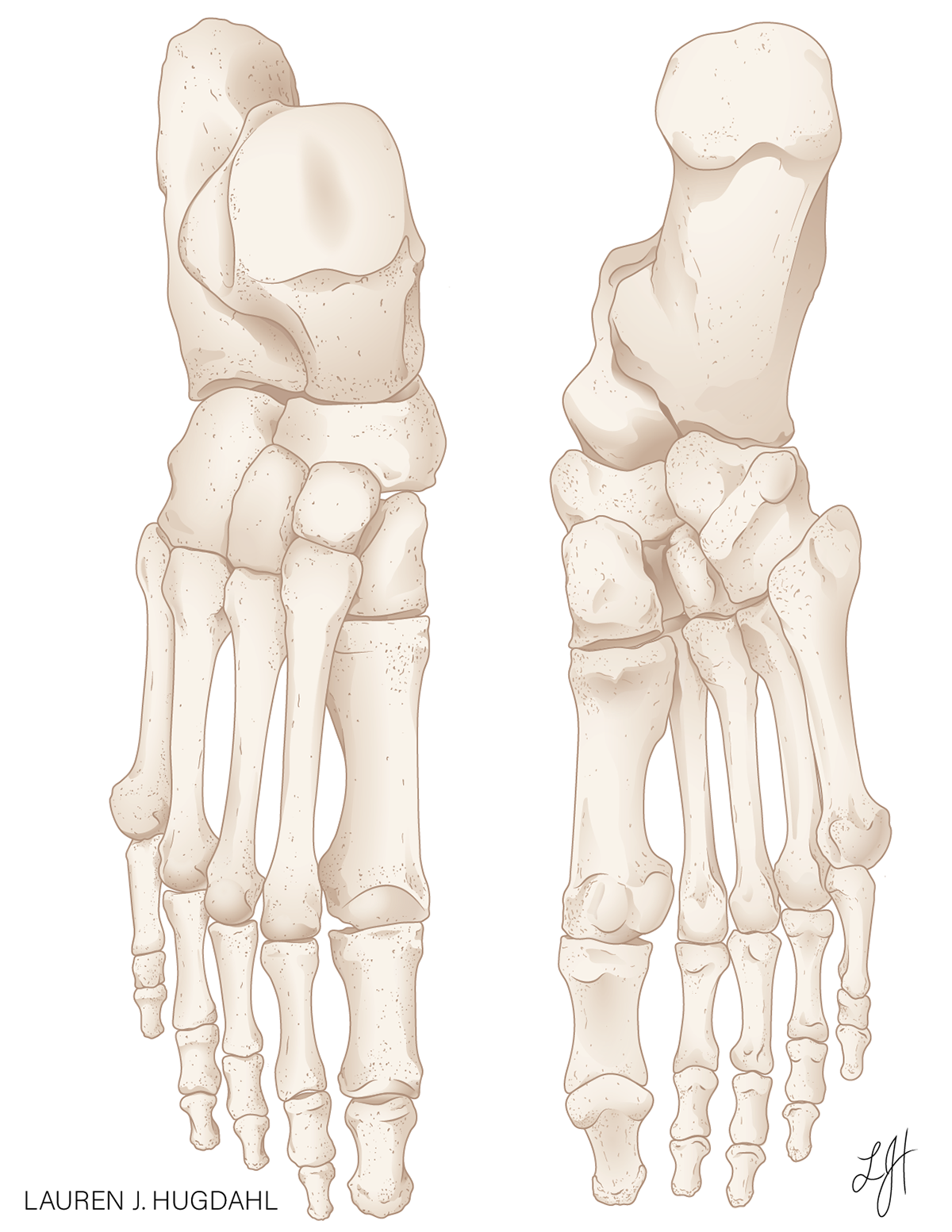
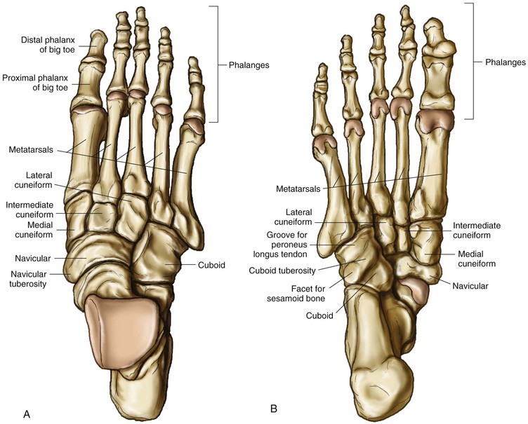
Tibia, fibula, talus, calcaneus, navicular, cuneiform, cuboid, distal phalanges, medial phalanges, proximal. Dorsiflexion occurs in both ankle joint and wrist joint. Arterial supply to the foot can be divided into plantar and dorsal components. The back of the hand or top of the foot. Each arises from a single metatarsal.
 Source: trialexhibitsinc.com
Plantar pressure patterns after rotationplasty resemble the plantar loading situation of the healthy foot with respect to area, force and loading time but reveal smaller peak pressure values whereas dorsal pressure patterns are characterised by a smaller loading area and decreased force but longer loading times, Borrowed from latin plantāre, present active infinitive of plantō. Arterial supply to the foot can be divided into plantar and dorsal components. The force was higher on the plantar. In humans the sole of the foot is anatomically referred to as the plantar aspect.
 Source: epainassist.com
The opposite side of the foot is called the plantar surface. Your toenails are on the dorsal side of the foot, because they are on the back (or upper) side of it. The lisfranc ligament is a strong band of tissue that connects the medial cuneiform to the base of the second metatarsal. What is the medical term for top of foot? The measurements were reproducible and indicated that the main loading areas of the rotated foot inside the prosthesis are medially on the dorsal aspect and in the heel and toe region or the heel and midfoot region on the plantar aspect.
 Source: behance.net
Dorsiflexion and plantar flexion are two motions of the body, decreasing the angle between two anatomical parts of the body. The plantar ligaments are stronger than those on the dorsal side (figure 12 & 13). In anatomy, the sole of the foot is called the plantar surface. Plantar vs dorsal what difference english. Anterior tibial artery, via dorsalis pedis and dorsal metatarsal arteries
 Source: custompilatesandyoga.com
Request pdf | plantar and dorsal foot loading measurements in patients after rotationplasty | the present study investigated the plantar and dorsal foot loading patterns inside the prosthesis of. The spur on the plantar surface of the calcaneus is duwe to chronic traction of the plantar fascia. The force was higher on the plantar. Plantar flexion is the lifting of the heel of the foot from the ground or pointing the toes downward. Medial process of calcaneal tuberosity.
 Source: myfootshop.com
In humans the sole of the foot is anatomically referred to as the plantar aspect. Medial process of calcaneal tuberosity. Each arises from a single metatarsal. Dorsiflexion and plantarflexion are special body movements involving the foot and ankle joint.during dorsiflexion, the angle between the dorsum of the foot a. Flexor digiti minimi brevis innervation.
 Source: easynotecards.com
Your toenails are on the dorsal side of the foot, because they are on the back (or upper) side of it. The opposite side of the hand is the palmar surface; Dorsiflexion occurs in both ankle joint and wrist joint. Arterial supply to the foot can be divided into plantar and dorsal components. As adjectives the difference between dorsal and plantar is that dorsal is (anatomy) with respect to, or concerning the side in which the backbone is located, or the analogous side of an invertebrate while plantar is pertaining to the bottom surface ( sole ) of the foot, as with plantar warts compare palmar.
 Source: cascadedafo.com
Dorsiflexion occurs in both ankle joint and wrist joint. Anatomy of plantar and dorsal foot bones related words: Borrowed from latin plantāre, present active infinitive of plantō. The top of the foot is called the dorsal of the foot because in anatomy the term dorsal refers to things which are on top or opposite the. The opposite side of the foot is called the plantar surface.
 Source: researchgate.net
Anterior tibial artery, via dorsalis pedis and dorsal metatarsal arteries The force was higher on the plantar. The dorsal loading area was smaller than the plantar area ( p =0.003). Borrowed from latin plantāre, present active infinitive of plantō. Each arises from a single metatarsal.
 Source: kenhub.com
What is the medical term for top of foot? Anterior tibial artery, via dorsalis pedis and dorsal metatarsal arteries In anatomy, the sole of the foot is called the plantar surface. The plantar interossei have a unipennate morphology, while the dorsal interossei are bipennate. The plantar and dorsal interossei comprise the fourth and final plantar muscle layer.
 Source: teachmeanatomy.info
Tibia, fibula, talus, calcaneus, navicular, cuneiform, cuboid, distal phalanges, medial phalanges, proximal. The opposite side of the hand is the palmar surface; The plantar and dorsal interossei comprise the fourth and final plantar muscle layer. The sole is the bottom of the foot. The spur on the plantar surface of the calcaneus is duwe to chronic traction of the plantar fascia.
 Source: doctorlib.info
From latin planta (“sole of the foot”). The top of the foot is called the dorsum of the foot. In humans the sole of the foot is anatomically referred to as the plantar aspect. From latin planta (“sole of the foot”). Request pdf | plantar and dorsal foot loading measurements in patients after rotationplasty | the present study investigated the plantar and dorsal foot loading patterns inside the prosthesis of.
 Source: musculoskeletalkey.com
Anterior tibial artery, via dorsalis pedis and dorsal metatarsal arteries Blood supply the vascularization of dorsal interossei muscles comes from several small arteries in the foot ; Borrowed from latin plantāre, present active infinitive of plantō. The dorsal loading area was smaller than the plantar area (p=0.003). Each arises from a single metatarsal.
This site is an open community for users to submit their favorite wallpapers on the internet, all images or pictures in this website are for personal wallpaper use only, it is stricly prohibited to use this wallpaper for commercial purposes, if you are the author and find this image is shared without your permission, please kindly raise a DMCA report to Us.
If you find this site adventageous, please support us by sharing this posts to your own social media accounts like Facebook, Instagram and so on or you can also save this blog page with the title plantar and dorsal foot by using Ctrl + D for devices a laptop with a Windows operating system or Command + D for laptops with an Apple operating system. If you use a smartphone, you can also use the drawer menu of the browser you are using. Whether it’s a Windows, Mac, iOS or Android operating system, you will still be able to bookmark this website.


