Your Plantar fascia anatomy images are available. Plantar fascia anatomy are a topic that is being searched for and liked by netizens now. You can Download the Plantar fascia anatomy files here. Get all royalty-free photos and vectors.
If you’re looking for plantar fascia anatomy images information related to the plantar fascia anatomy topic, you have pay a visit to the ideal site. Our website always gives you hints for refferencing the maximum quality video and picture content, please kindly search and find more enlightening video content and graphics that match your interests.
Plantar Fascia Anatomy. The subcutaneous tissue lying deep to the dorsal skin is loose and prone to oedema. This is evident also analyzing the anatomical terminology. Supporting the arch along the bottom (plantar) side of the foot between the heel bone (calcaneus, to be precise) and toes stretches a thick band of connective tissue called the plantar fascia. We are looking at how to identify the 3 bands of the plantar fascia and which band is most important in plantar fasciitis.
 Ideal Balance Center for Sports Medicine Acupuncture From idealbalanceacupuncture.blogspot.com
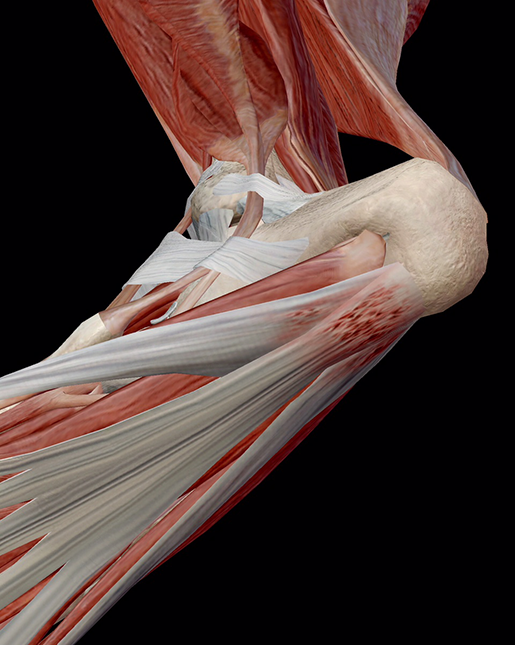
Analyzing its possible connections to the sural structures showed that this fascia is more closely connected to the paratenon of achilles tendon than to the achilles tendon, through the periosteum of the heel. It runs forward to the base of the toes and forms the medial arch on the foot. The plantar fascia is a thickened fibrous aponeurosis that originates from the medial tubercle of the calcaneus, runs forward to insert into the deep, short transverse ligaments of the metatarsal heads, dividing into 5 digital bands at the metatarsophalangeal joints and continuing forward to form the fibrous flexor sheathes on the plantar aspect of the toes. This is an anatomical dissection of the plantar fascia. The pf is a tissue firmly joined to plantar muscles and skin. On the foot, there are superficial (subcutaneous) and deep fasciae.
Although usually not disabling, plantar
Lateral band the reason you need to know this is because together the plantar fascia covers a large area of the foot and not. Follow the plantar fascia distally towards the toes. Because of its combined static and dynamic role in longitudinal arch support in the foot ( , 2 , , 3 ) and the capability of allowing the loading capacity on the foot during weight bearing, abnormalities of the pa are commonly encountered in the diagnostic. Fascia and septa divide other soft and bony tissues of the leg into three fascial compartments, namely, anterior, lateral, and posterior compartments. Nothing is known about its content of elastic fibers, the features of the extracellular matrix or the extent of innervation. The pf is a tissue firmly joined to plantar muscles and skin.
 Source: youtube.com
Although the plantar fascia (pf) has been studied quite well from a biomechanical viewpoint, its microscopic properties have been overlooked: Plantaris gets its name because in many mammals it inserts into the plantar aponeurosis. Supporting the arch along the bottom (plantar) side of the foot between the heel bone (calcaneus, to be precise) and toes stretches a thick band of connective tissue called the plantar fascia. Analyzing its possible connections to the sural structures showed that this fascia is more closely connected to the paratenon of achilles tendon than to the achilles tendon, through the periosteum of the heel. In the nomina anatomica only the term plantar aponeurosis is used to indicate this structure, but it is inserted in the chapter ‘fasciae’.
 Source: sportspodiatry.com.au
The plantar fascia is a thickened fibrous aponeurosis that originates from the medial tubercle of the calcaneus, runs forward to insert into the deep, short transverse ligaments of the metatarsal heads, dividing into 5 digital bands at the metatarsophalangeal joints and continuing forward to form the fibrous flexor sheathes on the plantar aspect of the toes. Assess longitudinally, with the probe on the plantar aspect of the heel. This dynamic mechanism can also be depicted as a triangle, with the plantar fascia represented by the hypotenuse of the triangle. On the foot, there are superficial (subcutaneous) and deep fasciae. Nonetheless, the superficial lamina presents some remarkable particularities at the level of the tarsus.
 Source: orthopaedicprinciples.com
The plantar fascia consists of thick connective tissue which supports the medial longitudinal arch on the sole (plantar side) of the foot. This article will cover the anatomy and functions of the plantaris muscle. In the nomina anatomica only the term plantar aponeurosis is used to indicate this structure, but it is inserted in the chapter ‘fasciae’. Plantar fasciitis is the result of collagen degeneration of the plantar fascia at the origin, the calcaneal tuberosity of the heel as well as the surrounding perifascial structures. It is a thick connective tissue, that functions to support and protect the underlying vital structures of the foot.
 Source: bettydroche.wordpress.com
The plantar fascia is a thickened fibrous aponeurosis that originates from the medial tubercle of the calcaneus, runs forward to insert into the deep, short transverse ligaments of the metatarsal heads, dividing into 5 digital bands at the metatarsophalangeal joints and continuing forward to form the fibrous flexor sheathes on the plantar aspect of the toes. This is evident also analyzing the anatomical terminology. In the various anatomical textbooks, the. The origin on the calcaneum encounters most of the weight bearing stress and is where plantar fascitis occurs. This is the 2nd video in our series on mri of the plantar fascia.
 Source: bodytechreview.com
This is an anatomical dissection of the plantar fascia. Supporting the arch along the bottom (plantar) side of the foot between the heel bone (calcaneus, to be precise) and toes stretches a thick band of connective tissue called the plantar fascia. However, in humans, the plantaris comes nowhere near it. It runs forward to the base of the toes and forms the medial arch on the foot. Ensure you sweep across through the entire width of the origin.
 Source: newtonrunning.com
The plantar fascia is a complex structure that extends from the medial calcaneal tubercle (the heel bone) to the proximal phalanges of the toes (the bone at the base of the toe) at the metatarsophalangeal (mtp) joints. Plantar fasciitis is the result of collagen degeneration of the plantar fascia at the origin, the calcaneal tuberosity of the heel as well as the surrounding perifascial structures. However, in humans, the plantaris comes nowhere near it. In the past, the plantar fascia has received more attention from the biomechanical engineers and clinicians than from anatomists. This contributes to optimal foot mobility and the spring of the foot.
 Source: ergonx.com.au
Arising predominantly from the calcaneal tuberosity, the plantar fascia attaches distally, through several slips, to the plantar aspect of the forefoot as well as the medial and lateral intermuscular septa. The fascia is thick centrally, known as aponeurosis and is thin along the sides. Supporting the arch along the bottom (plantar) side of the foot between the heel bone (calcaneus, to be precise) and toes stretches a thick band of connective tissue called the plantar fascia. It runs forward to the base of the toes and forms the medial arch on the foot. The plantar fascia runs from the calcaneus to the phalanges.
 Source: visiblebody.com
However, in humans, the plantaris comes nowhere near it. It is a thick connective tissue, that functions to support and protect the underlying vital structures of the foot. In the past, the plantar fascia has received more attention from the biomechanical engineers and clinicians than from anatomists. This is the 3rd video in our series on mri of the plantar fascia. The pf is a tissue firmly joined to plantar muscles and skin.
 Source: hubpages.com
Anatomy of the plantar fascia. On the plantar surface, the subcutaneous. Although the plantar fascia (pf) has been studied quite well from a biomechanical viewpoint, its microscopic properties have been overlooked: This is the 3rd video in our series on mri of the plantar fascia. The plantar fascia, or plantar aponeurosis, forms part of the deep fascia of the sole of the foot and provides a strong mechanical linkage between the calcaneus and the toes.
 Source: stefanduell.tumblr.com
Follow the plantar fascia distally towards the toes. This article will cover the anatomy and functions of the plantaris muscle. This dynamic mechanism can also be depicted as a triangle, with the plantar fascia represented by the hypotenuse of the triangle. This is an anatomical dissection of the plantar fascia. As the muscle crosses both the knee and ankle joints, it weakly assists with knee and plantar flexion.
 Source: americanfoot.com
Together, they are called the plantar fascia ligaments and are surrounded. Plantaris gets its name because in many mammals it inserts into the plantar aponeurosis. This article will cover the anatomy and functions of the plantaris muscle. The plantar aponeurosis is the modification of deep fascia, which covers the sole. The plantar fascia plays an important role in the normal biomechanics of the foot.
 Source: dr7physioandpod.com.au
Anatomy of the plantar fascia. Normal bands today’s post is again a video on mri of the plantar fascia anatomy. As the palmar fascia of the hand, it has two laminas, a superficial one and a deep one, whose disposition varies according to the species. However, in humans, the plantaris comes nowhere near it. This article will cover the anatomy and functions of the plantaris muscle.
 Source: saragerow.com
The plantar fascia is a thick band of tissue that is inserted to the under surface of the heel bone (calcaneus). The plantar fascia plays an important role in the normal biomechanics of the foot. Plantaris gets its name because in many mammals it inserts into the plantar aponeurosis. However, in humans, the plantaris comes nowhere near it. This is an anatomical dissection of the plantar fascia.
Source: idealbalanceacupuncture.blogspot.com
This article will cover the anatomy and functions of the plantaris muscle. The fascia is thick centrally, known as aponeurosis and is thin along the sides. Fascia and septa divide other soft and bony tissues of the leg into three fascial compartments, namely, anterior, lateral, and posterior compartments. On the plantar surface, the subcutaneous. In the past, the plantar fascia has received more attention from the biomechanical engineers and clinicians than from anatomists.
 Source: vaidyaveekshan.blogspot.com
Because of its combined static and dynamic role in longitudinal arch support in the foot ( , 2 , , 3 ) and the capability of allowing the loading capacity on the foot during weight bearing, abnormalities of the pa are commonly encountered in the diagnostic. Arising predominantly from the calcaneal tuberosity, the plantar fascia attaches distally, through several slips, to the plantar aspect of the forefoot as well as the medial and lateral intermuscular septa. In the past, the plantar fascia has received more attention from the biomechanical engineers and clinicians than from anatomists. In order for patients and professionals to understand plantar fasciitis, they must have at least a simple appreciation of the anatomy of the plantar fascia. The plantar fascia is a complex structure that extends from the medial calcaneal tubercle (the heel bone) to the proximal phalanges of the toes (the bone at the base of the toe) at the metatarsophalangeal (mtp) joints.
 Source: pinterest.ca
In the various anatomical textbooks, the. As the muscle crosses both the knee and ankle joints, it weakly assists with knee and plantar flexion. This is evident also analyzing the anatomical terminology. Lateral band the reason you need to know this is because together the plantar fascia covers a large area of the foot and not. The plantar aponeurosis is the modification of deep fascia, which covers the sole.
 Source: floridaortho.com
Because of its combined static and dynamic role in longitudinal arch support in the foot ( , 2 , , 3 ) and the capability of allowing the loading capacity on the foot during weight bearing, abnormalities of the pa are commonly encountered in the diagnostic. On the plantar surface, the subcutaneous. The plantar fascia (fascia plantaris) is stronger and more complex than the dorsal fascia. The plantar fascia is a thick band of tissue that is inserted to the under surface of the heel bone (calcaneus). It runs from the medial tuberosity of the calcaneus (the inside of the heel bone) forward to the heads of the metatarsal bones.
 Source: hybridpainrelief.com
The plantar fascia is a complex structure that extends from the medial calcaneal tubercle (the heel bone) to the proximal phalanges of the toes (the bone at the base of the toe) at the metatarsophalangeal (mtp) joints. The plantar fascia is a thickened fibrous aponeurosis that originates from the medial tubercle of the calcaneus, runs forward to insert into the deep, short transverse ligaments of the metatarsal heads, dividing into 5 digital bands at the metatarsophalangeal joints and continuing forward to form the fibrous flexor sheathes on the plantar aspect of the toes. In the various anatomical textbooks, the. Not to be confused with heel spurs, a different injury with similar symptoms, plantar fasciitis occurs when the plantar fascia becomes irritated and sore. Plantar fascia 10 according to stecco et al.
This site is an open community for users to do sharing their favorite wallpapers on the internet, all images or pictures in this website are for personal wallpaper use only, it is stricly prohibited to use this wallpaper for commercial purposes, if you are the author and find this image is shared without your permission, please kindly raise a DMCA report to Us.
If you find this site adventageous, please support us by sharing this posts to your favorite social media accounts like Facebook, Instagram and so on or you can also bookmark this blog page with the title plantar fascia anatomy by using Ctrl + D for devices a laptop with a Windows operating system or Command + D for laptops with an Apple operating system. If you use a smartphone, you can also use the drawer menu of the browser you are using. Whether it’s a Windows, Mac, iOS or Android operating system, you will still be able to bookmark this website.