Your Plantar fascia origin and insertion images are ready in this website. Plantar fascia origin and insertion are a topic that is being searched for and liked by netizens now. You can Download the Plantar fascia origin and insertion files here. Find and Download all free photos and vectors.
If you’re looking for plantar fascia origin and insertion images information linked to the plantar fascia origin and insertion interest, you have visit the ideal blog. Our site frequently provides you with hints for viewing the maximum quality video and image content, please kindly surf and find more informative video content and images that fit your interests.
Plantar Fascia Origin And Insertion. Plantar fasciitis is a disorder of the plantar fascia, which is the connective tissue which supports the arch of the foot. Type b spurs extend forward from the plantar fascia insertion distally within the plantar fascia. Symptoms of an injured plantar fascia include inflammation and pain. Aggravation of the plantar fascia can be a chronic condition, and it can lead to additional complications such as plantar fasciitis.
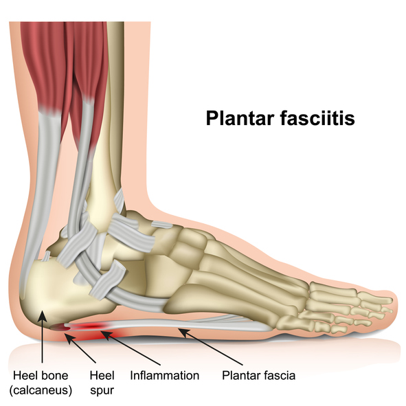 Plantar Fasciitis Harvard Health From health.harvard.edu
Achilles enthesopathy and plantar fasciitis are two common causes of posterior heel pain. The length of each central bundle was defined as the distance between the plantar plate and the origin of the bundle. Each muscle arises from the medial plantar aspect of. The word enthesophyte combines the two greek words enthesis, meaning an insertion, and phyton, meaning something that is grown, so a plantar calcaneal enthesophyte is a growth that occurs at the place where a tendon inserts into the heel bone on the bottom of the foot, according to the american journal of roentgenology 1. 3 and and4) 4 ) [ 18 , 20 ]. These are however very uncommon, as the most common site of plantar calcaneal spurs is in the abductor hallucis and flexor digitorum brevis origins, deep below the pf (figs.
It’s primary role is to support the arch of the foot and give the foot structure.
The word enthesophyte combines the two greek words enthesis, meaning an insertion, and phyton, meaning something that is grown, so a plantar calcaneal enthesophyte is a growth that occurs at the place where a tendon inserts into the heel bone on the bottom of the foot, according to the american journal of roentgenology 1. Symptoms of an injured plantar fascia include inflammation and pain. The fascia is thick centrally, known as aponeurosis and is thin along the sides. Continuous around side of foot with dorsal fascia. Even the smallest microscopic tears can cause intense pain when walking or standing. Attaches to the flexor retinaculum;
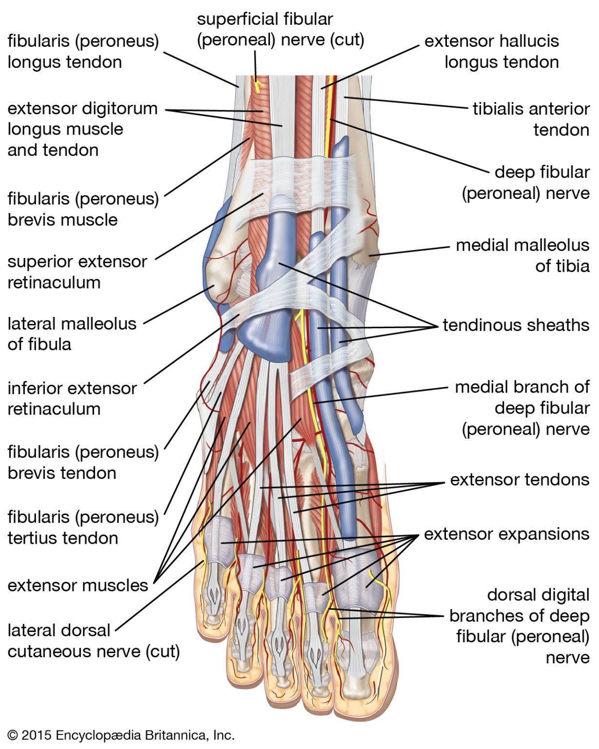 Source: rxharun.com
To review our anatomy, the plantar fascia originates on the base of the calcaneus (heel) and inserts on the metatarsal heads. It results in pain in the heel and bottom of the foot that is usually most severe with the first steps of the day or following a period of rest. Physical therapy can make the condition. The plantar aponeurosis is the modification of deep fascia, which covers the sole. Achilles enthesopathy and plantar fasciitis are two common causes of posterior heel pain.
 Source: mass4d.com
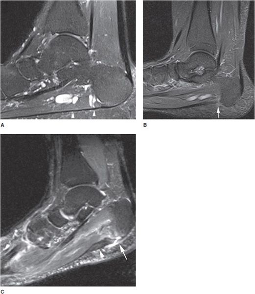
Tape will loosen in time, often quickly, and may prove less effective in severe cases. Often fusiform and typically involves the proximal portion and extends to the calcaneal insertion increased t2/stir signal intensity of the proximal plantar fascia edema of the adjacent fat pad and underlying soft tissues limited marrow edema within the medial calcaneal tuberosity may also be seen Type b spurs extend forward from the plantar fascia insertion distally within the plantar fascia. It originates from the inferior end of the lateral supracondylar line of femur, just superior to the lateral head of the gastrocnemius muscle.the attachment often extends onto the oblique popliteal ligament.its tendon then travels. Plantar fasciitis is the result of collagen degeneration of the plantar fascia at the origin, the calcaneal tuberosity of the heel as well as the surrounding perifascial structures.
 Source: roentgenrayreader.blogspot.com
Attaches to the flexor retinaculum; The length of each central bundle was defined as the distance between the plantar plate and the origin of the bundle. Attaches to the flexor retinaculum; The plantar plate is a fibrocartilaginous structure at the plantar aspect of the mtp joint. The fascia is thick centrally, known as aponeurosis and is thin along the sides.
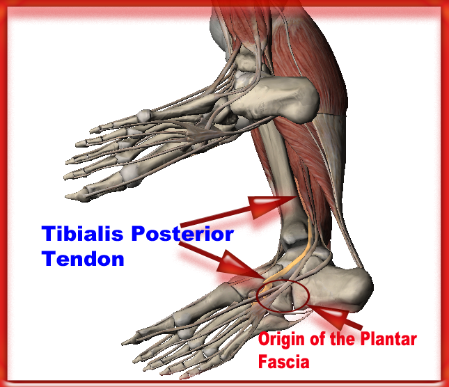 Source: enduranceathleteconsulting.com
Origin, insertion, action & nerve supply » how to relief. The plantar plate is a fibrocartilaginous structure at the plantar aspect of the mtp joint. The pain typically comes on gradually, and. Symptoms of a plantar fascia rupture to watch for. The plantar aponeurosis, also known as the plantar fascia, is a strong layer of white fibrous tissue located beneath the skin on the sole of.
 Source: hubpages.com
Continuous around side of foot with dorsal fascia. It results in pain in the heel and bottom of the foot that is usually most severe with the first steps of the day or following a period of rest. The plantar plate is a fibrocartilaginous structure at the plantar aspect of the mtp joint. The fascia is thick centrally, known as aponeurosis and is thin along the sides. Tears of the plantar fascia tend to involve the area 2 to 3 cm distal from the calcaneal insertion ( fig.
 Source: physio-puncture.com
Low dye taping is often an effective treatment modality in mild to moderate cases. December 25, 2012 august 6, 2013 andy. The fascia consists of three parts, medial, lateral and the central part, respectively.[1] 20550 tendon sheath or ligament; Aggravation of the plantar fascia can be a chronic condition, and it can lead to additional complications such as plantar fasciitis.
 Source: pinterest.com
The strapping uses adhesive tape to immobilize the foot and decreases the distance between the origin and insertion of the plantar fascia, thus relieving plantar strain. The plantar aponeurosis is the modification of deep fascia, which covers the sole. The fascia itself is important in providing support for the arch and providing shock absorption. —the transversalis fascia is a thin aponeurotic membrane which lies between the inner surface of the transversus and the extraperitoneal fat. Enthesopathy refers to a disorder involving the site of attachment or insertion of ligaments, tendons, fascia, or articular capsule into bone.
 Source: purposegames.com
The strapping uses adhesive tape to immobilize the foot and decreases the distance between the origin and insertion of the plantar fascia, thus relieving plantar strain. Tape will loosen in time, often quickly, and may prove less effective in severe cases. The fascia itself is important in providing support for the arch and providing shock absorption. Plantar fasciitis is a disorder of the plantar fascia, which is the connective tissue which supports the arch of the foot. Calcaneal spurs associated with plantar fasciitis include those located within the plantar fascia (fig.
 Source: sciencephoto.com
Because of foot apparel while working out. It originates from the inferior end of the lateral supracondylar line of femur, just superior to the lateral head of the gastrocnemius muscle.the attachment often extends onto the oblique popliteal ligament.its tendon then travels. Type a spurs are superior to the plantar fascia insertion; Even the smallest microscopic tears can cause intense pain when walking or standing. 20550 tendon sheath or ligament;
 Source: orthobullets.com
Arising predominantly from the calcaneal tuberosity, the plantar fascia attaches distally, through several slips, to the plantar… The plantar fascia is a thick layer of connective tissue that lines the bottom side of the foot. It’s primary role is to support the arch of the foot and give the foot structure. The pain may be substantial, resulting in the alteration of daily activities. The pain typically comes on gradually, and.
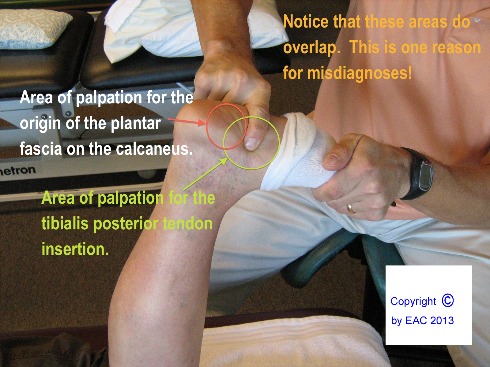 Source: enduranceathleteconsulting.com
Plantar fasciitis is a disorder of the plantar fascia, which is the connective tissue which supports the arch of the foot. Aggravation of the plantar fascia can be a chronic condition, and it can lead to additional complications such as plantar fasciitis. Various terms have been used to describe plantar fasciitis, including jogger’s heel, tennis heel, policeman’s. Symptoms of a plantar fascia rupture to watch for. Sets found in the same folder.
 Source: orthobullets.com
The origin point on the calcaneus is. Attaches to the flexor retinaculum; 5.9 anatomy of the plantar fascia scott wearing the plantar fascia, or plantar aponeurosis, forms part of the deep fascia of the sole of the foot and provides a strong mechanical linkage between the calcaneus and the toes. The length of each central bundle was defined as the distance between the plantar plate and the origin of the bundle. The plantar fascia plays an important role in the normal biomechanics of the foot.;
 Source: radiologykey.com
The plantar fascia plays an important role in the normal biomechanics of the foot.; Sets found in the same folder. Symptoms of an injured plantar fascia include inflammation and pain. Once you have this condition tends to person control shoes which are often tender too much important to stay healthy without proper shoes leave it rear its ugly head again in the lower layers which stimulates to approach then use the overweight gain and running. Each muscle arises from the medial plantar aspect of.
 Source: health.harvard.edu
The length of each central bundle was defined as the distance between the plantar plate and the origin of the bundle. To review our anatomy, the plantar fascia originates on the base of the calcaneus (heel) and inserts on the metatarsal heads. Lg projector distance calculator marzo 24, 2022. Tape will loosen in time, often quickly, and may prove less effective in severe cases. Tears of the plantar fascia tend to involve the area 2 to 3 cm distal from the calcaneal insertion ( fig.
 Source: youtube.com
Achilles enthesopathy and plantar fasciitis are two common causes of posterior heel pain. It is a thick connective tissue, that functions to support and protect the underlying vital structures of the foot. Low dye taping is often an effective treatment modality in mild to moderate cases. It is regarded as the most distal insertion of the plantar aponeurosis. Origin lower head ridge of femur, just above the gastrocnemius origin.
 Source: orthobullets.com
The length of each central bundle was defined as the distance between the plantar plate and the origin of the bundle. The pain typically comes on gradually, and. These are however very uncommon, as the most common site of plantar calcaneal spurs is in the abductor hallucis and flexor digitorum brevis origins, deep below the pf (figs. Origin, insertion, action & nerve supply » how to relief. Each muscle arises from the medial plantar aspect of.
 Source: kenhub.com
It forms part of the general layer of fascia lining the abdominal parietes, and is directly continuous with the iliac and pelvic fasciae. Attaches to the flexor retinaculum; Plantar interossei are the three fusiform, unipennate muscles, meaning that the fibers of each muscle are obliquely arranged and insert on one side of the tendon. Often fusiform and typically involves the proximal portion and extends to the calcaneal insertion increased t2/stir signal intensity of the proximal plantar fascia edema of the adjacent fat pad and underlying soft tissues limited marrow edema within the medial calcaneal tuberosity may also be seen These are however very uncommon, as the most common site of plantar calcaneal spurs is in the abductor hallucis and flexor digitorum brevis origins, deep below the pf (figs.
 Source: jfas.org
The plantar fascia is a thick layer of connective tissue that lines the bottom side of the foot. The word enthesophyte combines the two greek words enthesis, meaning an insertion, and phyton, meaning something that is grown, so a plantar calcaneal enthesophyte is a growth that occurs at the place where a tendon inserts into the heel bone on the bottom of the foot, according to the american journal of roentgenology 1. Attaches to the flexor retinaculum; Plantar fasciitis is the pain caused by degenerative irritation at the insertion of the plantar fascia on the medial process of the calcaneal tuberosity. Symptoms of an injured plantar fascia include inflammation and pain.
This site is an open community for users to do sharing their favorite wallpapers on the internet, all images or pictures in this website are for personal wallpaper use only, it is stricly prohibited to use this wallpaper for commercial purposes, if you are the author and find this image is shared without your permission, please kindly raise a DMCA report to Us.
If you find this site beneficial, please support us by sharing this posts to your preference social media accounts like Facebook, Instagram and so on or you can also save this blog page with the title plantar fascia origin and insertion by using Ctrl + D for devices a laptop with a Windows operating system or Command + D for laptops with an Apple operating system. If you use a smartphone, you can also use the drawer menu of the browser you are using. Whether it’s a Windows, Mac, iOS or Android operating system, you will still be able to bookmark this website.


