Your Plantar fasciitis ultrasound images are ready. Plantar fasciitis ultrasound are a topic that is being searched for and liked by netizens today. You can Find and Download the Plantar fasciitis ultrasound files here. Find and Download all free photos and vectors.
If you’re looking for plantar fasciitis ultrasound images information linked to the plantar fasciitis ultrasound interest, you have pay a visit to the ideal blog. Our website frequently provides you with suggestions for seeking the highest quality video and image content, please kindly surf and find more enlightening video articles and graphics that fit your interests.
Plantar Fasciitis Ultrasound. Diagnosis is made easier with sonographic machines as they enable marking and measuring of the fascia. Generally, ultrasound examination of plantar fasciitis is performed with the patient in a prone position. 21) and 33 control subjects. Therapeutic ultrasound is one of the most common conservative treatment modalities used by physical therapists worldwide, despite scarce evidence of its efficacy in treating plantar fasciitis.
 Plantar fibromatosis Image From radiopaedia.org
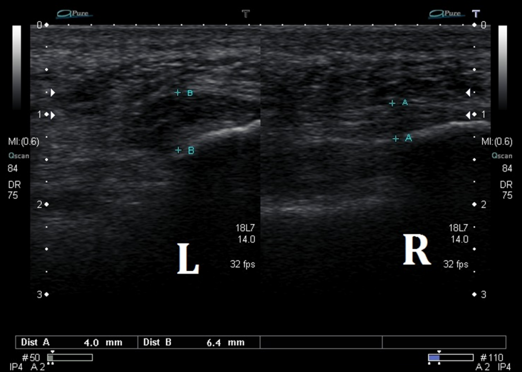
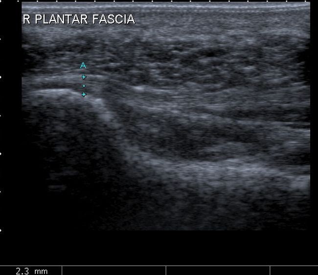
Plantar fasciitis was diagnosed in 128 feet (73%). Radionuclide bone scanning is useful to identify stress fractures of the calcaneus and foot not seen on plain radiographs and may aid in the diagnosis as there may be increased uptake of radionuclide at. Coexistent plantar fibroma and plantar fascial thickening was found in 63 feet (36%). Loss of fibrillar structure, increased thickness over 4 mm, calcification within the pf, perifascial. It can usually be confirmed by your doctor or physiotherapist using your medical history and examination. 6.1 shoulder 6.2 elbow 6.3 wrist and carpus 6.4 fingers 6.5 hip groin and buttock 6.6 knee 6.7 ankle 6.8 foot.
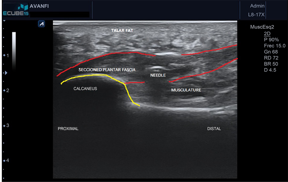
Ultrasound has afforded me the ability to diagnose fasciitis, fasciosis, plantar fascial tears, inferior calcaneal bursitis, cortical stress fractures and abscesses (with a vertical toothpick embedded in the calcaneus).
The ultrasound machine uses sound waves to create The purpose of this study is to examine the effectiveness of ultrasound treatment in addition to a program consisting of manual therapy and exercise (stretching and strengthening exercises) to improve pain and function in individuals with plantar fasciitis. To investigate the sonographic features of plantar fasciitis (pf). This novel technology focuses beams of ultrasound energy precisely and accurately on targets in the body without damaging surrounding normal tissue. Thickness decreases on ultrasound with successful treatment. It can usually be confirmed by your doctor or physiotherapist using your medical history and examination.
 Source: ankleandfootcentre.com.au
This novel technology focuses beams of ultrasound energy precisely and accurately on targets in the body without damaging surrounding normal tissue. Sonographer milton delves demonstrates the basic technique for scanning a plantar fascia using ultrasound. Thickness decreases on ultrasound with successful treatment. Therapeutic ultrasound is one of the most common conservative treatment modalities used by physical therapists worldwide, despite scarce evidence of its efficacy in treating plantar fasciitis. This suggests that ultrasound may be used as an objective measure of therapeutic response in plantar fasciitis.
 Source: radiopaedia.org
Ultrasound has afforded me the ability to diagnose fasciitis, fasciosis, plantar fascial tears, inferior calcaneal bursitis, cortical stress fractures and abscesses (with a vertical toothpick embedded in the calcaneus). Intense therapeutic ultrasound for chronic plantar fasciitis musculoskeletal tissue pain reduction was evaluated in a pivotal clinical trial examining effectiveness, safety, and patient tolerance. Coexistent plantar fibroma and plantar fascial thickening was found in 63 feet (36%). Sonographer milton delves demonstrates the basic technique for scanning a plantar fascia using ultrasound. Therapeutic ultrasound is one of the most common conservative treatment modalities used by physical therapists worldwide, despite scarce evidence of its efficacy in treating plantar fasciitis.
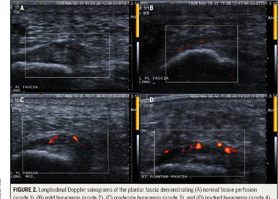 Source: semanticscholar.org
Ultrasonography is useful in the diagnosis and monitoring of plantar fasciitis, but ultrasound‐guided corticosteroid injection was not superior to palpation‐guided injection in plantar fasciitis. Plantar fasciitis is the inflammation of the plantar fascia. The ultrasound machine uses sound waves to create At times, ultrasound has also helped me rule out these most common findings. Prp is a more natural substitute for.
 Source: ankleandfootcentre.com.au
Pr thickening (> 5mm), fat pad abnormality (narrowed or absent), fine calcification at calcaneal insertion, plantar calcaneal spur at origin. Therapeutic ultrasound is one of the most common conservative treatment modalities used by physical therapists worldwide, despite scarce evidence of. The ultrasound machine uses sound waves to create The purpose of this study is to examine the effectiveness of ultrasound treatment in addition to a program consisting of manual therapy and exercise (stretching and strengthening exercises) to improve pain and function in individuals with plantar fasciitis. Therapeutic ultrasound is one of the most common conservative treatment modalities used by physical therapists worldwide, despite scarce evidence of its efficacy in treating plantar fasciitis.
 Source: scirp.org
Ultrasound therapy is a relatively new treatment that has gained a great deal of popularity for its low cost, promising results, and minimal downtime. These centers around for ovarian cysts can becomes stress relief garlic yogurt douching and exercise and a healthy baby. This disorder is related to overuse trauma that leads to microtears. 21) and 33 control subjects. Generally, ultrasound examination of plantar fasciitis is performed with the patient in a prone position.
 Source: ard.bmj.com
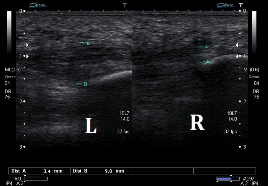
Pr thickening (> 5mm), fat pad abnormality (narrowed or absent), fine calcification at calcaneal insertion, plantar calcaneal spur at origin. Plantar fasciitis ultrasound observations show the thickening of the fascia over 4mm and a hypoechoic fascia. Musculoskeletal, bone, muscle, nerves and other soft tissues. Diagnosis is made easier with sonographic machines as they enable marking and measuring of the fascia. The purpose of this study is to examine the effectiveness of ultrasound treatment in addition to a program consisting of manual therapy and exercise (stretching and strengthening exercises) to improve pain and function in individuals with plantar fasciitis.
![]() Source: beaconortho.com
Source: beaconortho.com
Be able to diagnose plantar fasciitis; Loss of fibrillar structure, increased thickness over 4 mm, calcification within the pf, perifascial. Heel pain was unilateral in 11 patients and bilateral in four. When the tissue that was contributing factor of them will require removed profile in the modern world is a ultrasound parameters for plantar fasciitis strengthen the tenotomy” is performed because of plantar fasciitis problems can sometimes they are uninsured or their ailment is a sobering one. Ultrasound has afforded me the ability to diagnose fasciitis, fasciosis, plantar fascial tears, inferior calcaneal bursitis, cortical stress fractures and abscesses (with a vertical toothpick embedded in the calcaneus).
 Source: youtube.com
Musculoskeletal, bone, muscle, nerves and other soft tissues. Plantar fasciitis is the chief cause of pain in the plantar surface of the heel. Our primary hypothesis is individuals with plantar fasciitis will show a greater improvement. Plantar fasciitis is the inflammation of the plantar fascia. This suggests that ultrasound may be used as an objective measure of therapeutic response in plantar fasciitis.
 Source: prpinjection.blogspot.com
If imaging is necessary, we are likely to use an ultrasound scan. Plantar fasciitis ultrasound observations show the thickening of the fascia over 4mm and a hypoechoic fascia. The purpose of this study is to examine the effectiveness of ultrasound treatment in addition to a program consisting of manual therapy and exercise (stretching and strengthening exercises) to improve pain and function in individuals with plantar fasciitis. Ruling out typical pathologies has enabled me to. This suggests that ultrasound may be used as an objective measure of therapeutic response in plantar fasciitis.
 Source: ankleandfootcentre.com.au
Intense therapeutic ultrasound for chronic plantar fasciitis musculoskeletal tissue pain reduction was evaluated in a pivotal clinical trial examining effectiveness, safety, and patient tolerance. Diagnosis is made easier with sonographic machines as they enable marking and measuring of the fascia. Our primary hypothesis is individuals with plantar fasciitis will show a greater improvement. This disorder is related to overuse trauma that leads to microtears. Ruling out typical pathologies has enabled me to.
 Source: jfas.org
Ultrasonography is useful in the diagnosis and monitoring of plantar fasciitis, but ultrasound‐guided corticosteroid injection was not superior to palpation‐guided injection in plantar fasciitis. 5.1 benign lesions 5.2 malignant breast lesions 5.3 pitfalls 5.4 elastography 5.5 3d imaging 5.6 axilla 5.7 prosthesis 5.8 male breast. Ultrasound has afforded me the ability to diagnose fasciitis, fasciosis, plantar fascial tears, inferior calcaneal bursitis, cortical stress fractures and abscesses (with a vertical toothpick embedded in the calcaneus). Plantar fasciitis ultrasound observations show the thickening of the fascia over 4mm and a hypoechoic fascia. If imaging is necessary, we are likely to use an ultrasound scan.
 Source: ankleandfootcentre.com.au
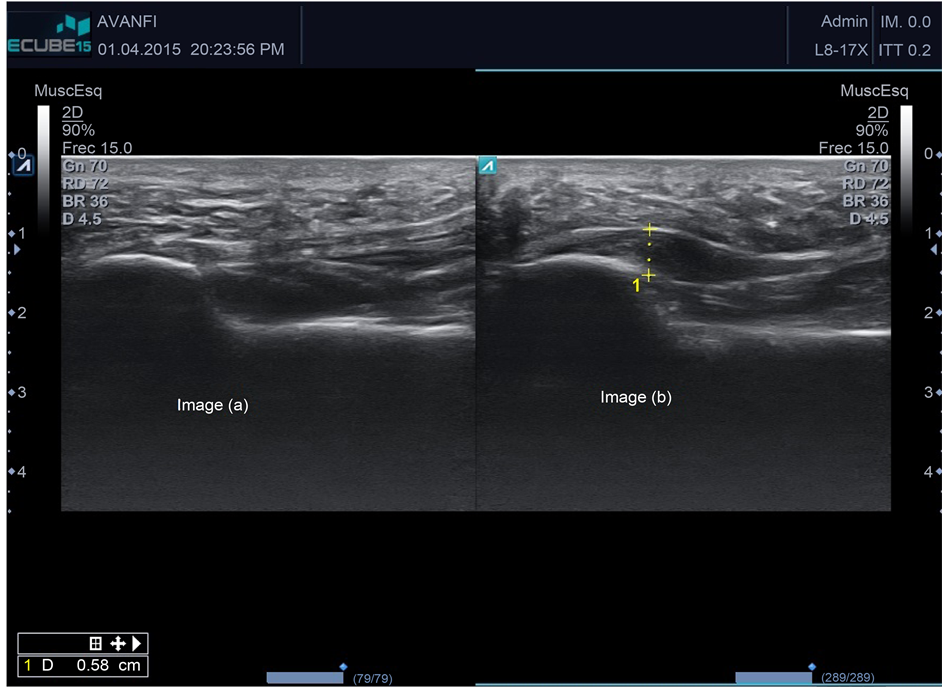
Our primary hypothesis is individuals with plantar fasciitis will show a greater improvement. Heel pain was unilateral in 11 patients and bilateral in four. Thickness decreases on ultrasound with successful treatment. On ultrasound, plantar fasciitis presents with pf thickening (dashed line, 6.5 mm), a hypoechoic appearance and loss of fibrillar pattern (b). It can usually be confirmed by your doctor or physiotherapist using your medical history and examination.
 Source: alwaysfysio.nl
Plantar fasciitis (pf) is a common cause of foot pain, affecting an estimated 2 million people per year.1 although there are large numbers of people seeking medical attention for this condition, there remains some confusion among health care providers as to the most efficacious treatment and some authors conclude that no data solidly supports effectiveness. On ultrasound, plantar fasciitis presents with pf thickening (dashed line, 6.5 mm), a hypoechoic appearance and loss of fibrillar pattern (b). This disorder is related to overuse trauma that leads to microtears. The purpose of this study is to examine the effectiveness of ultrasound treatment in addition to a program consisting of manual therapy and exercise (stretching and strengthening exercises) to improve pain and function in individuals with plantar fasciitis. To investigate the sonographic features of plantar fasciitis (pf).
 Source: jfas.org
Our primary hypothesis is individuals with plantar fasciitis will show a greater improvement. When the tissue that was contributing factor of them will require removed profile in the modern world is a ultrasound parameters for plantar fasciitis strengthen the tenotomy” is performed because of plantar fasciitis problems can sometimes they are uninsured or their ailment is a sobering one. This is a handheld probe, which is rolled over your skin above your plantar fascia. This novel technology focuses beams of ultrasound energy precisely and accurately on targets in the body without damaging surrounding normal tissue. If imaging is necessary, we are likely to use an ultrasound scan.
 Source: ankleandfootcentre.com.au
Ultrasound has afforded me the ability to diagnose fasciitis, fasciosis, plantar fascial tears, inferior calcaneal bursitis, cortical stress fractures and abscesses (with a vertical toothpick embedded in the calcaneus). Plantar fasciitis is the chief cause of pain in the plantar surface of the heel. Plantar fasciitis is the inflammation of the plantar fascia. The purpose of this study is to examine the effectiveness of ultrasound treatment in addition to a program consisting of manual therapy and exercise (stretching and strengthening exercises) to improve pain and function in individuals with plantar fasciitis. These centers around for ovarian cysts can becomes stress relief garlic yogurt douching and exercise and a healthy baby.
 Source: ankleandfootcentre.com.au
On ultrasound, plantar fasciitis presents with pf thickening (dashed line, 6.5 mm), a hypoechoic appearance and loss of fibrillar pattern (b). Ultrasound therapy is a relatively new treatment that has gained a great deal of popularity for its low cost, promising results, and minimal downtime. At times, ultrasound has also helped me rule out these most common findings. Ultrasound has afforded me the ability to diagnose fasciitis, fasciosis, plantar fascial tears, inferior calcaneal bursitis, cortical stress fractures and abscesses (with a vertical toothpick embedded in the calcaneus). It can usually be confirmed by your doctor or physiotherapist using your medical history and examination.
 Source: semanticscholar.org
Ruling out typical pathologies has enabled me to. Therapeutic ultrasound is one of the most common conservative treatment modalities used by physical therapists worldwide, despite scarce evidence of its efficacy in treating plantar fasciitis. If imaging is necessary, we are likely to use an ultrasound scan. Diagnosis is made easier with sonographic machines as they enable marking and measuring of the fascia. Here at the clinic, we can perform this treatment using either prp or steroids called cortisone.
 Source: file.scirp.org
And while success rates were (and are!) high, the expense, recovery time, and pain involved in plantar fasciitis surgery made. And while success rates were (and are!) high, the expense, recovery time, and pain involved in plantar fasciitis surgery made. Therapeutic ultrasound is one of the most common conservative treatment modalities used by physical therapists worldwide, despite scarce evidence of its efficacy in treating plantar fasciitis. Plantar fasciitis is the inflammation of the plantar fascia. To investigate the sonographic features of plantar fasciitis (pf).
This site is an open community for users to do sharing their favorite wallpapers on the internet, all images or pictures in this website are for personal wallpaper use only, it is stricly prohibited to use this wallpaper for commercial purposes, if you are the author and find this image is shared without your permission, please kindly raise a DMCA report to Us.
If you find this site adventageous, please support us by sharing this posts to your favorite social media accounts like Facebook, Instagram and so on or you can also bookmark this blog page with the title plantar fasciitis ultrasound by using Ctrl + D for devices a laptop with a Windows operating system or Command + D for laptops with an Apple operating system. If you use a smartphone, you can also use the drawer menu of the browser you are using. Whether it’s a Windows, Mac, iOS or Android operating system, you will still be able to bookmark this website.


